ECG of the week #4: short of breath with wide complex tachycardia
Test yourself with this week’s ECG case. Then scroll down for a structured step-by-step interpretation and clinical pearl.
Clinical Scenario
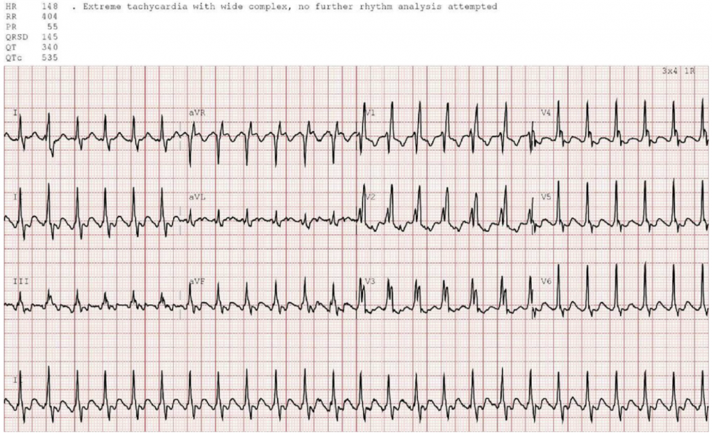
A 75 year old presented with shortness of breath. The computer interpretation is “Extreme tachycardia with wide complex, no further rhythm analysis attempted.”
ECG
Question
Attempt rhythm analysis.
ECG Analysis
H: wide complex tachy@150 with inferior inverted flutter waves@300
E: 2:1 conduction, typical RBBB (rsR’ in V1, rapid R wave and slow s wave in I)
A: normal axis
R: early R from RBBB
T: normal voltages
S: flutter waves obscure ST/T
Answer
atrial flutter with 2:1 and RBBB
Clinical Pearl
Regular wide complex tachycardia without P waves is monomorphic VT until proven otherwise. But when there are typical flutter and typical BBB morphology this can be identified as atrial flutter with BBB