Test yourself with this week’s ECG case. Then scroll down for a structured step-by-step interpretation and clinical pearl.
Clinical Scenario
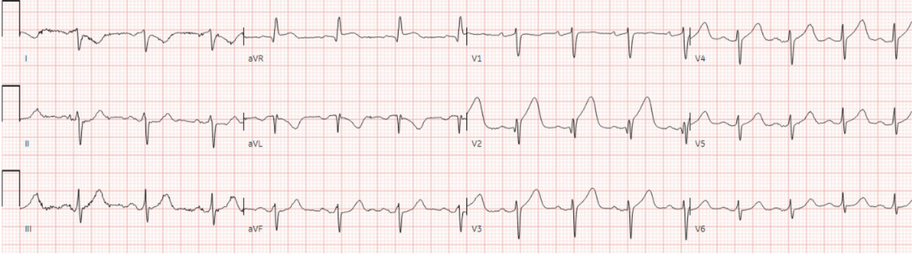
A previously health 60 year old presented with acute chest pain
ECG
Question
Any signs of occlusion, and if so where is the culprit?
ECG Analysis
H: normal sinus
E: normal conduction
A: normal axis
R: normal R-wave progression
T: normal voltages
S: STE V4-5, hyperacute T waves V2-5 and inferior with reciprocal disproportionate TWI in aVL
Answer
Wraparound or distal LAD occlusion
Clinical Pearl
Distal or wraparound LAD occlusion can cause STE/hyperacute T waves across the precordium and into the inferior leads