SUBSCRIBE for Upcoming Course Announcements
Test yourself with this week’s ECG case. Then scroll down for a structured step-by-step interpretation and clinical pearl.
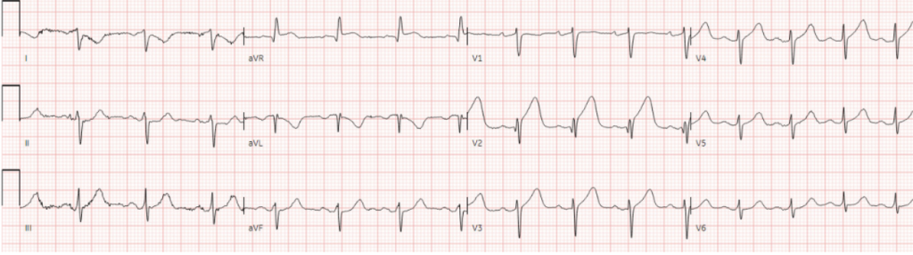
A 70 year-old presented with acute chest pain.
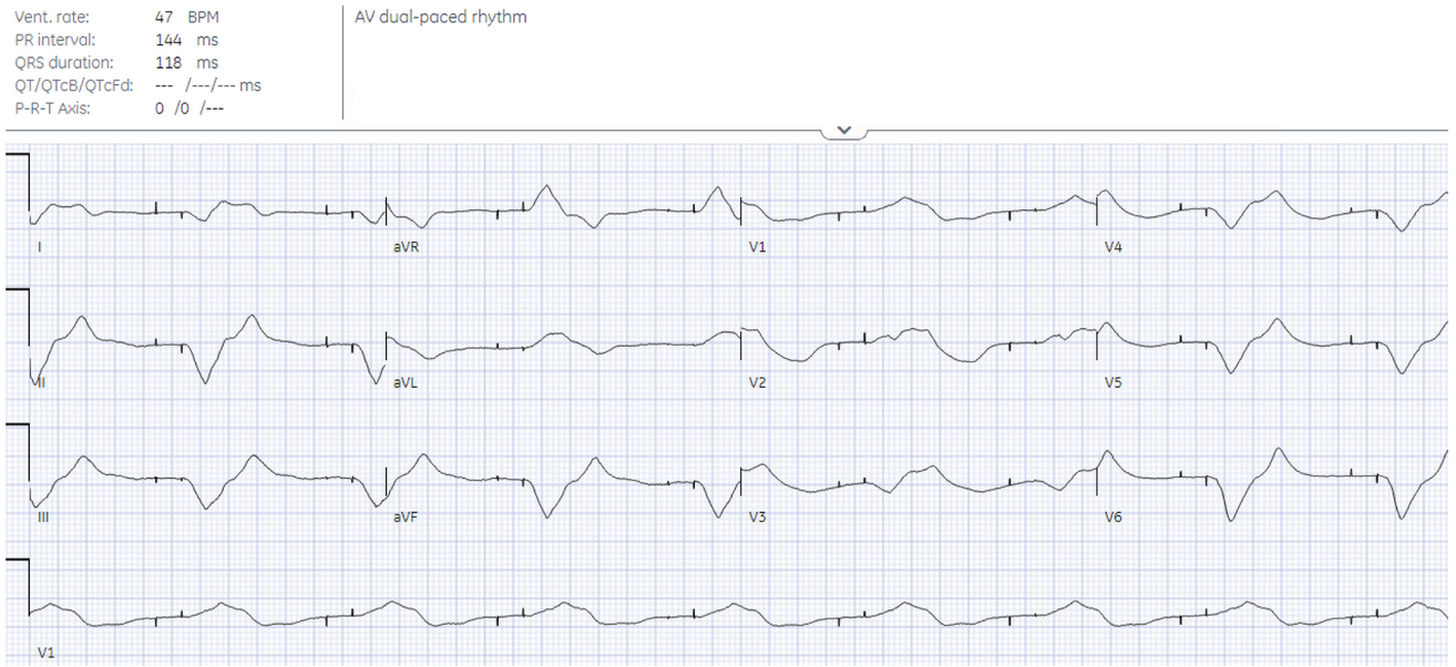
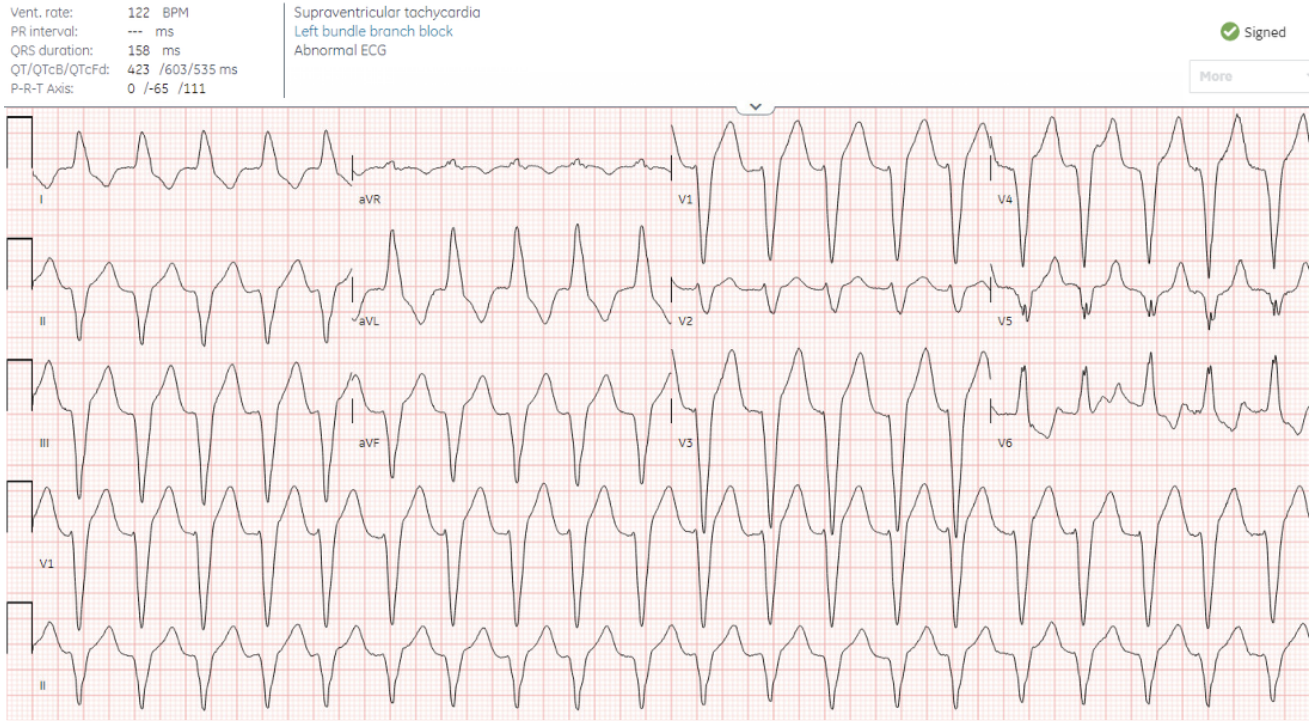
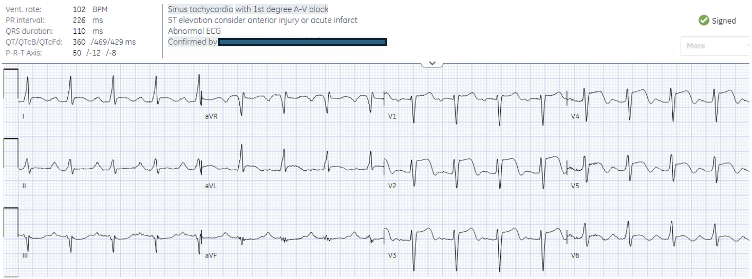
There are a number of ECG abnormalities – but which are real and which are fake?
H: normal sinus in II but P inverted in I/upright in aVR (lead reversal)
E: normal
A: right axis with pseudo-Q waves from RA/LA reversal (inverted P-QRS-T in I/aVL/aVR)
R: small Q in V2 and late progression
T: normal
S: STE/hyperacute T V2-5, aVR (actual aVL) and inverted in I, and STD in III (actual II)
1. pseudo-lateral Q waves from right arm/left arm lead reversal
2. real proximal LAD occlusion
1. Sinus rhythm with normal lead placement has upright P in II>I and inverted in aVR. RA/LA reversal has inverted P/QRS/T in I, aVL/aVR are fliped and III/II are flipped.
2. Hyperacute T waves (broad based, look inflated relative to QRS) can help identify occlusion MI when there is minimal STE