SUBSCRIBE for Upcoming Course Announcements
Test yourself with this week’s ECG case. Then scroll down for a structured step-by-step interpretation and clinical pearl.
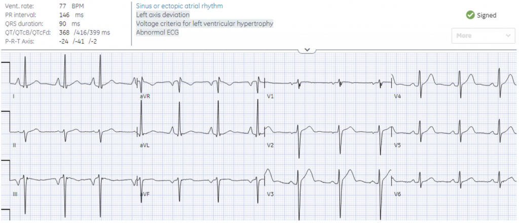
A 25 year old presented with shortness of breath.
What’s the ECG abnormality?
Heart rate/rhythm: sinus rhythm but P wave I>II and inverted P/QTS/T in III = LA/LL reversal
Electrical conduction: normal intervals
Axis: left axis from lead reversal
R-wave progression: normal
Tall/small voltages: pseudo-LVH from lead reversal
ST/T: normal
Left arm/left leg reversal, no LVH
Sinus rhythm with normal limb lead placement has P wave upright in II > I, negative in aVR. There are many types of limb lead reversal–including left arm/left leg which can be identified by P wave II > I and inverted P/QRS/T in III. This results in I/II switching places, aVF/aVL switching places, and III inverted.