ECG of the week #13: 65 year old with 13 hours of chest pain – is this a ‘late presenting STEMI’?
Test yourself with this week’s ECG case. Then scroll down for a structured step-by-step interpretation and clinical pearl.
Clinical Scenario
A 65 year old presented with 13 hours of chest pain
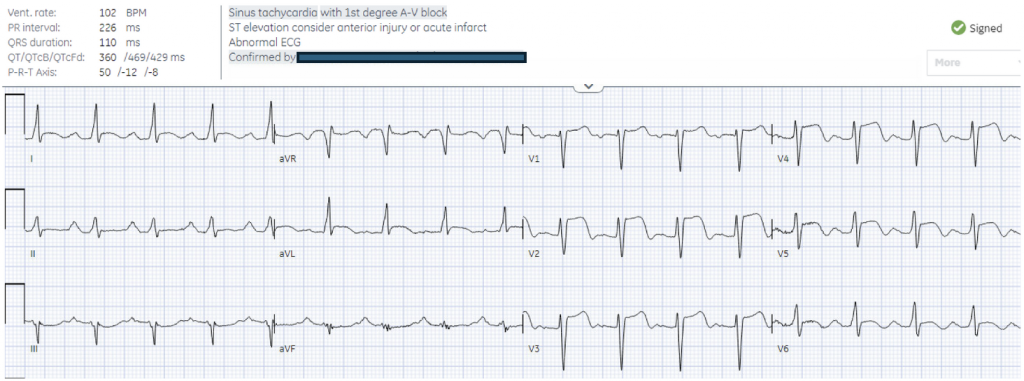
ECG
Question
Based on symptoms, this is a ‘late presenting STEMI'(>12 hours), which is traditionally considered to benefit less from reperfusion because of myocardium has already been lost. But does the ECG show a subacute or acute occlusion?
ECG Analysis
H: sinus borderline tachy
E: first degree AV block
A: normal axis
R: delayed but no Q waves
T: normal voltages
S: anterolateral STE with mild terminal T wave inversion, reciprocal inferior STD
Answer
Acute anterolateral STEMI without Q waves. Cath: 100% LAD occlusion.
First hs-troponin I was only 700ng/L (correlating with the ECG rather than the symptoms), peak 130,000
Clinical Pearl
Prolonged symptoms does not mean prolonged occlusion, as there can be intermittent reperfusion and reocclusion. The ECG can be a better indicator of occlusion time: from acute (no Q waves + STE + hyperacute T waves) to subacute (Q waves + shallow convex STE + T wave inversion).